

Incidental findings in radiology can be a cause for concern for patients, their doctors, and their families. It is important to understand what these findings are and what they may mean for your health (for patients) or the health of your patients (for providers). In this blog post, I will discuss the definition of incidental findings, common types of incidental findings, and what you need to know about them. I will also provide tips on how to best manage some of the most common incidental findings.
What is an Incidental Finding?
An incidental finding is as its name describes: a finding that was not the main focus of a radiology exam and was found by chance. Incidental findings can be further broken down into two types: determinate findings (the findings are diagnostic of an entity – we know what it is) and indeterminate (a finding is made, but the imaging features are not diagnostic, frequently requiring additional testing to obtain a diagnosis – we don’t know what it is). These unrelated findings may be of clinical importance, such as incidental cancer requiring treatment, or of no clinical importance, such as a benign renal cyst (a sac of fluid that can safely be ignored).
Incidental tumors are typically the most concerning and anxiety-provoking unrelated findings we make and can be either benign (non-cancerous) or malignant (cancerous). Benign tumors can be safely ignored while malignant tumors require treatment or surveillance.
Anytime a person is imaged, there’s a chance of making an incidental finding. And whenever a radiologist looks at an examination, we not only try to address the clinical problem at hand (such as pain) but also try to identify anything else that could prove problematic in the future. We want to identify malignant cancers while they’re curable and identify pathological processes to help guide management and prevent bad outcomes in the future. For example, identifying coronary calcium indicative of coronary artery disease in a young individual allows for a change in lifestyle and starting heart-protective medications to prevent or delay a future heart attack. This can be a defining moment in a patient’s life.
The scope of a radiologist is not only to address the problem at hand but also to identify anything of potential future significance while being mindful of the costs of associated follow-up testing. This is one important area where we add value in medicine.
Prevalence of Incidental Findings
Prevalence of incidental findings varies by imaging modality (ultrasound, CT, MRI, etc.) and the body part being imaged. Several meta-analyses exist (in a meta-analysis numerous research articles are reviewed and data is pooled to give greater confidence in the results) evaluating the prevalence with a recent study in the BMJ from 2018 reporting the following incidental finding rates:
- Brain MRI – 22%
- Spine MRI – 22%
- Chest CT – 45%
- Cardiac MRI – 34%
- CT Colonography (Virtual Colonoscopy) – 38%
In this list, you can see that we frequently discover incidental findings on all kinds of exams.
The prevalence of malignant incidental findings was 11% across all studies (11% of incidental findings consisted of a malignant tumor) with breast, ovarian, renal, thyroid, colon, and prostate cancers most commonly discovered. It should be no surprise that this list consists of many of the most common cancers that people are familiar with.
Common Types of Incidental Findings
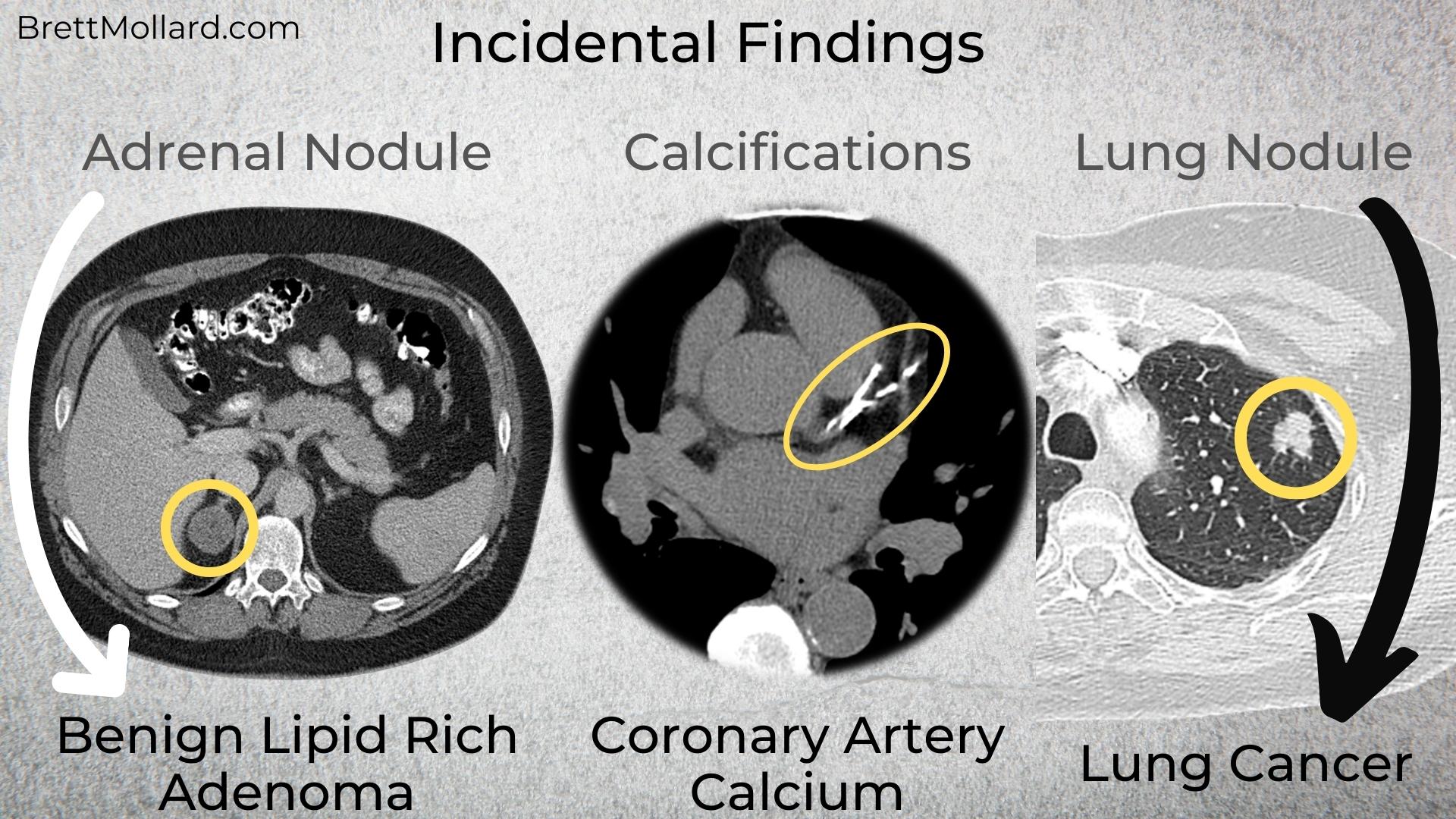
Many different types of incidental findings can be found in imaging studies including benign and malignant tumors and other pathological processes. Some of the more common ones include:
Atherosclerotic Plaque:
- Atherosclerotic plaque is a buildup of fatty deposits in the arteries and is a common finding on CTs of all body parts. They can calcify over time.
Calcifications:
- Calcifications are small calcium deposits that can be found in various organs on a CT scan. They can occur in stones (example: kidney stones, gallstones), be associated with atherosclerotic plaque (coronary artery disease), and be seen with various other processes.
Cysts:
- Cysts are fluid-filled sacs that can be found in any organ of the body. Cysts contain simple fluid and are nearly always of no clinical significance unless the cyst is so large that it pushes on adjacent structures. Cysts are incredibly common and frequently found in kidneys and livers.
Lymph Nodes:
- Lymph nodes are small, kidney bean-shaped structures that are part of the lymphatic system. They can be seen on ultrasound, mammograms, CT, or MRI exams.
Tumors:
- Tumors can be benign (non-cancerous) or malignant (cancerous). They can be found in any organ and are most often seen on ultrasound, CT, or MRI.
While not an exhaustive list, some of the most commonly encountered incidental findings are covered in the next sections.
Circulatory System (Blood Vessels)
Coronary Arteries:
Coronary artery calcifications are one of the most common and most important incidental findings that we make. Risks of coronary artery disease vary depending on the amount of coronary calcium, patient age, gender, and race. Higher amounts of coronary artery calcium are associated with higher levels of cardiac events and mortality related to coronary artery disease (CAD) such as heart attacks. When discovered, patients can undergo risk factor assessment, health and lifestyle changes, and be treated with various medications depending on patient risk factors and test results.
Aorta and Major Branch Arteries:
The aorta – the largest of the blood vessels supplying blood to the entire body – can have atherosclerotic plaque (calcified and/or non-calcified) and can form aneurysms (where it is dilated larger than normal). Ascending thoracic aortic aneurysms (where the aorta comes off of the heart in the chest) and abdominal aortic aneurysms are at increased risk of rupturing (bursting) over time, which is typically fatal. With risks including instant death, an aortic aneurysm is a “must make” diagnosis that can save lives. Large aortic aneurysms typically undergo imaging surveillance once detected and may require surgery for definitive treatment after they reach a certain size threshold.
All major branch arteries arising from the aorta can have significant narrowing or even be occluded, from the carotid arteries supplying the brain to the femoral arteries supplying the legs. Carotid artery disease, for example, can lead to stroke.
Lungs and Legs:
Pulmonary emboli (blood clots in the lungs) and deep venous thromboses (DVTs; blood clots in the legs) are uncommon inciden